BRAINatomy 1
Optimising cognition in children treated with brain radiotherapy

Introduction to the challenges of treating paediatric brain cancers with Radiotherapy
Brain tumours are the second most common childhood cancer (after leukaemia), with just over 400 children with brain tumours diagnosed in the UK every year. About three out of four children with brain tumours survive at least five years following their diagnosis, with over 70% of children surviving long term (defined as more than five years but largely equates to more for paediatric brain tumours). Although most children are cured of their brain cancer, the treatment methods can cause complications for a lot of patients.
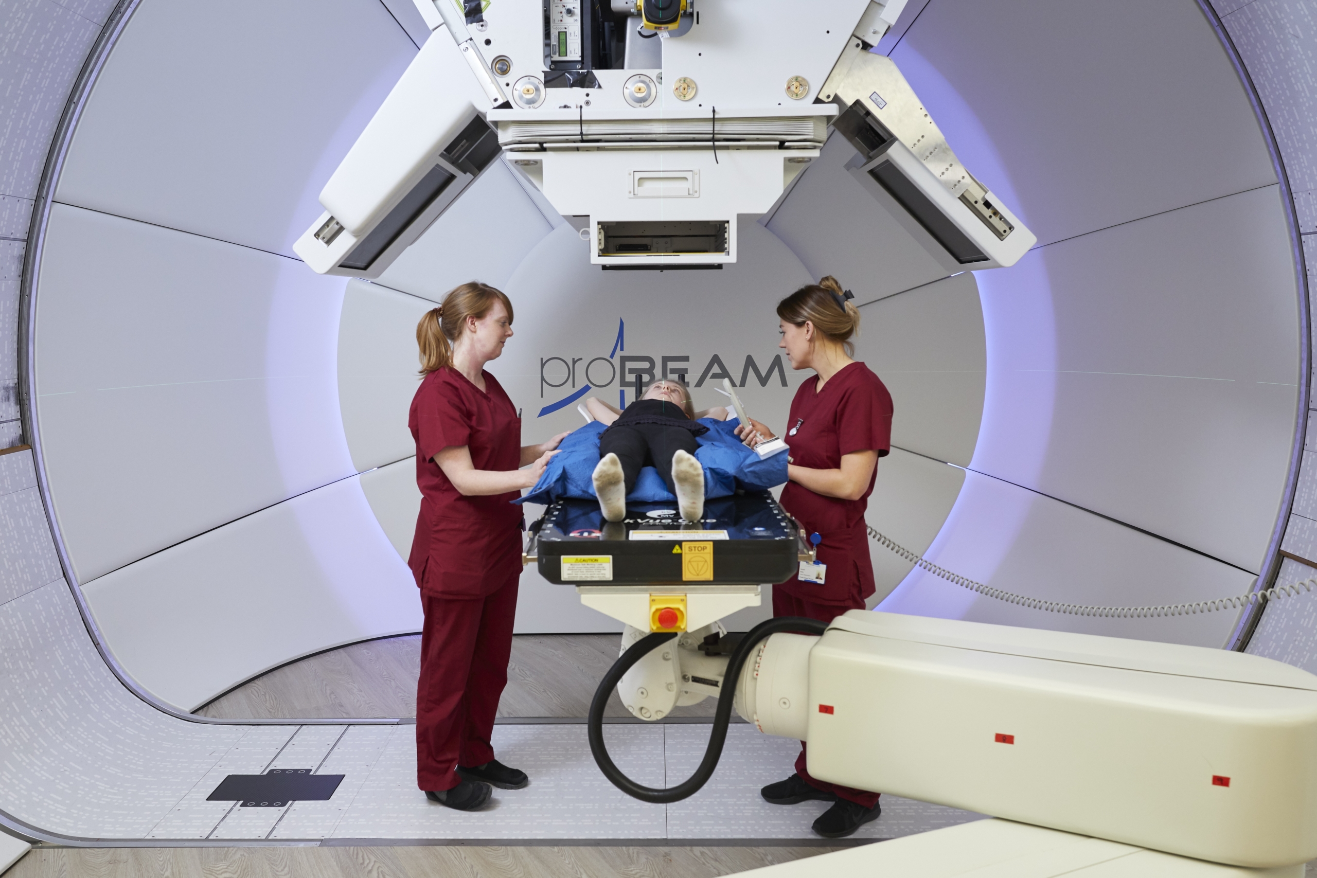
Radiotherapy, a high-precision cancer treatment which uses radiation beams (x-rays or protons), is a key component of curative treatment for many paediatric brain tumours. However, despite improvements in the targeting of the radiation, radiotherapy can damage functional brain subunits and is responsible for observed changes in brain function in children. Survivors can experience lifelong side-effects, such as poor memory or hormonal disruption, affecting their ability to work and overall quality-of-life.
There have been previous limitations in lack of understanding around which functional brain units contribute most to late toxicity, alongside an absence of risk prediction models. This knowledge gap has been a limiting factor in improving radiotherapy and maximising patient benefits from recent advances such as Proton Beam Therapy (PBT).
“BRAINatomy shows how we can use large “real-world” datasets to learn how to develop kinder treatments. Thanks to new methodologies such as machine learning, radiomics and image-based data mining, we can analyse much larger datasets than we ever did, retaining detailed information about each patient’s scans and treatment to work out why some children have very poor learning outcomes while others don’t, and using knowledge we gain about the biological effects of radiotherapy on normal brain to limit the damage we cause.”
Professor Martin McCabe
University of Manchester and Honorary Consultant Paediatric Oncologist, The Christie NHS Foundation Trust

Background of BRAINatomy 1
To understand how to reduce the harm of treatment, a team of world-leading doctors, biologists, physicists, patients and patient advocates were brought together to address this challenge. Led by The University of Manchester in collaboration with The Christie NHS Foundation Trust, the Royal Manchester Children’s Hospital and international collaborators at St Jude Children’s Research Hospital (USA) and the University of Groningen (Netherlands), and funded through a Stand Up To Cancer®-Cancer Research UK Paediatric Cancer New Discoveries Challenge grant, the BRAINatomy project was established to improve the outcomes for children diagnosed with brain tumours.
The inception of this study in 2021 was spearheaded by Dr Martin McCabe of the University of Manchester, Honorary Consultant Paediatric Oncologist at The Christie.
The Manchester solution
The initial aim of this study was to understand the origins of brain damage in children receiving treatment for brain tumours by identifying factors at diagnosis that correlated with later impairments. This was done by determining specific brain regions where radiation dose correlated with brain and hormonal damage, and by studying the biological effects on individual brain cells of conventional radiotherapy and proton therapy.
A cohort of about 250-300 patients – approximately 200 treated at St Jude Children’s Research Hospital, USA, and 100 treated at The Christie – were studied throughout the course of this project. By analysing baseline MRI scans in patients newly diagnosed with brain tumours, the team identified regions where abnormal scans before treatment correlated with later deficits in learning and hormone production.
Through image-based data mining (IBDM) methods, the team also identified subunits within the brain where the dose of radiotherapy received correlated with observed changes in brain function. This work provided an ‘atlas’, i.e. a database of areas of the brain to specifically avoid during radiotherapy to minimise treatment side-effects.
Finally, the team identified significant inflammation of some parts of the brain after radiotherapy, and differences in the biological effects of proton therapy and conventional radiotherapy in different brain compartments.

The change in standards of care
Using these groundbreaking new methods and data from hundreds of children, the team found regions of the brain that appear to be sensitive to damage at the time of diagnosis, and regions where radiation dose is especially harmful to the growing brain. They are now testing whether we can avoid those regions using modern radiotherapy technology. This could be done, for example, by using advanced treatments such as Proton Beam Therapy or changing the angles of the radiation beams to avoid the most sensitive regions of the brain. This work could then inform the next generation of clinical trials involving cranial radiotherapy.
In addition to providing information about how to safely change radiotherapy planning practice, the programme’s experimental approach has also given complementary information to identify children at increased risk of cognitive damage because of the dose they received to the identified “sensitive regions” and take action to rectify this.
Finally, BRAINatomy investigators has clarified that the hypothalamus, the part of the brain that controls all other hormone glands, might play a specific role in mediating cognitive damage, developing the pre-clinical evidence and laying the groundwork for innovative therapeutic strategies like microglial depletion therapy. The clinical impacts of these approaches, if successful, will ultimately be measured in fewer learning disabilities, greater independence and better quality of life in childhood brain tumour survivors.
The future of the research project and a look ahead to BRAINatomy 2
The work done so far looks at children who have been treated with conventional radiotherapy. This is now being expanded to include children treated with Proton Beam Therapy (PBT) in the UK, the USA, and the Netherlands.
As the BRAINatomy project progresses, it holds the promise of transforming the landscape of paediatric brain tumour treatments. The researchers’ goal is to identify brain regions sensitive to radiotherapy, enabling the development of tailored treatments that mitigate long-term cognitive and endocrine issues. The potential creation of a prediction model could revolutionise decision-making for clinicians, offering a tool to optimise survival while minimising adverse effects.
To help future children avoid some of the damaging effects of treatment, our researchers are now testing in the current phase of research whether identifying hormone deficiencies early and giving drugs to reduce the level of inflammation in the brain can improve the lives of children once they’ve finished treatment for their cancers.

Meet the Team
Team & Work-Package Leads
Martin McCabe – Team & WP Lead (University of Manchester)
Tom Merchant – Co-Team & WP Lead (St Jude Children’s Research Hospital)
Lara Barazzuol – Team Principal & WP Lead (University Medical Center Groningen)
Stavros Stivaros – WP Lead (University of Manchester)
Marianne Aznar – WP Lead (University of Manchester)
Marcel van Herk – WP Lead (University of Manchester)
Project Manager
Kate Vaughan – PM (Manchester)
Co-Investigators
Peter Clayton – (University of Manchester)
Gareth Price– (University of Manchester)
Early Career Investigators
Abigail Bryce-Atkinson (University of Manchester)
Angie Davey (University of Manchester)
Simon Dockrell (University of Manchester)
Eliana Vasquez Osorio (University of Manchester)
Lydia Wilson (St Jude Children’s Research Hospital)
Fakriddin Pirlepesov (St Jude Children’s Research Hospital)
Jorden Cunningham (St Jude Children’s Research Hospital)
Luiza Reali-Nazario (University Medical Center Groningen)
Daniel Vasquez (University Medical Center Groningen)
Daniela Voshart (University Medical Center Groningen)
Data Manager
Lesley Albutt (University of Manchester)
Patients & Advocates
James Adams – Patient
Helen Bulbeck – Advocate
Joshua Goddard – Patient
Adam Thompson – Advocate
Funders
BRAINatomy is headed by the University of Manchester in collaboration with the St Jude Children’s Research Hospital (USA), the University of Groningen (The Netherlands), The University of Birmingham (UK), Princess Máxima Center for Paediatric Oncology (The Netherlands)
It is funded by Stand Up To cancer (SU2C) and Cancer Research UK through the SU2C – CRUK Pediatric Cancer New Discoveries Challenge along with support from braintrust for patient involvement and engagement.
Organisations
The University of Manchester, The Christie NHS Foundation Trust, Manchester University NHS Foundation Trust
International collaborators
St Jude Children’s Research Hospital (Memphis, USA), University Medical Center, Groningen (The Netherlands, The University of Birmingham (UK), Princess Máxima Center for Pediatric Oncology (The Netherlands)
Main contact names
Dr Martin McCabe (lead) / Professor Marianne Aznar (WP lead)


